The Australian Organoid Facility is a not-for-profit automation facility, providing quality-assured organoids for basic, translational and contract research.
Our goal is to help Queensland researchers access organoids reliably and affordably to accelerate 3D biological research in Australia.
We use state-of-the-art automation technologies to produce organoids at low cost. We are also equipped to optimally develop new organoid systems at scale and to perform sophisticated high-content analysis on complex 3D biological systems.
We provide a reliable pipeline of organoids, which can complement pre-clinical studies for drug candidates or assist the development of biomaterials and micro-engineered biosystems.
Why organoids?
3D culturing is superior to more traditional 2D culturing because it encapsulates tissue complexity, greater cellular interactions and signalling, and reduces the need for animal models that can be expensive and time consuming.
Organoids are self-organised 3D tissues derived from stem cells or clinical samples. They can provide more robust examination of tissue physiology, disease modelling, and drug screening.
Contact us
Get in touch to learn more about our research.
Doctor Nathan Godde
Facility Manager
aof@aibn.uq.edu.au
Doctor Maneet Bhatia
Research Officer
aof@aibn.uq.edu.au
Find us on social media
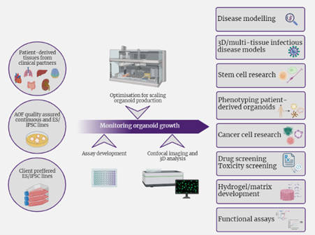 Our Services
- High throughput multi-tissue organoid production.
- Organoid model, optimisation and upscaling services.
- High throughput functional/drug screening.
- High content imaging and analysis.
- High throughput nucleic acid extraction.
- Collaborative development of new and innovative organoid models.
- Consulting: support for grants- budgeting, project development.
- Technical support.
The Australian Organoid Facility uses quality-assured continuous and stem cell lines and large batches of Matrigel and Cultrex to minimise batch-to-batch variations.
Facilities and Equipment
The Australian Organoid Facility (AOF) is located at The University of Queensland in the Australian Institute for Bioengineering and Nanotechnology.
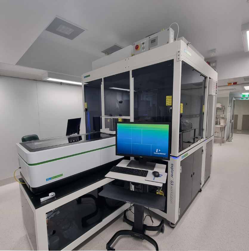
We use a custom designed, state-of-the-art automation platform called the AOF Explorer G3 Work Station (Revity) to produce, screen and analyse organoids.
It is located in a dedicated cleanroom and maintains a sterile environment through a HEPA filtration system.
The Explorer G3 has three levels, the main deck, a middle and a lower deck used to house various instrumentation and equipment.
The workstation has a track system plate handler robotic arm that integrates equipment across the decks facilitating regular media changes and observations without manual intervention.
Equipment includes:
Cell Explorer G3 Workstation (Revity)
Inside the Cell explorer G3 Workstation (Revvity)
- JANUS™ automated liquid hander
- 8-tip Varispan head provides independent variable sampler probe spacing for multi-tipped processing
- Modular Dispense Technology (MDT) head used for stamping 96 samples at once
- High-content imaging systems
- Opera Phenix™ Plus spinning disk confocal system
- Celigo whole-well image cyclometer
- Others
- Liconic automated incubators: 37OC and 4OC
- plate::handler™ FLEX 750 robotic arm
chemagic™ 360 automated nucleic acid purification system
Brain organoids
Cerebral organoids (COs) are 3-dimensional in vitro models with a cellular composition and structural organisation that is representative of the developing human brain.
COs are generated using a version of the protocol outlined by Lancaster, 2022 and modified for automation. This procedure can be further modified to generate several other region-specific brain organoids upon request.
Applications:
- Study of normal and pathological brain development.
- Modelling neurological disorders.
- Drug development.
- Biomaterials development.
Kidney organoids
Kidney organoids (KOs) are generated using a modified version of the Friedman et al.(2015), protocol and form self-organising 3D structures containing functional renal cell types.
KOs can recapitulate cellular and structural aspects of developing nephrons.
Potential applications:
- Modelling kidney diseases.
- Development studies.
- Nephrotoxicity screenings.
Blood vessel organoids
Supply of O2 and nutrients can limit the development of organoids in vitro. Therefore, remodelling of functional blood vessel networks is important.
Derived from hPSCs, blood vessel organoids (BVO) generated using the protocols of Wimmer et al (2019) produce functional blood vessels with a lumen and basement membrane deposition.
Potential applications:
- Study of vascular diseases.
- Study of organ development/transplantation.
- Drug testing.
- Study of tumour angiogenesis.
- Blood vessel organoids incorporated with other organoids to increase functionality. For example, vascularised kidney organoids, vascularised liver organoids.
Cancer Spheroids/Organoids
Cancer Spheroids are simple multi-cellular models that represent a more in vivo-like tumor tissue incorporating the tumor oxygen gradient and cellular interactions.
Potential applications:
- Therapeutic studies.
- Preclinical drug discovery/testing.
- Study of cell-cell and cell-matrix interactions.
We can expand and analyse a range of patient-derived cancer organoids that are researcher sourced or obtained by us through commercial suppliers such as Cellesce.
Resources:
- Large batches of GFR-Matrigel, Cultrex and ATCC-BME.
- Established cancer spheroid models for two breast cancer cell lines- MDA-MB-231 and MCF7 and one colon cancer cell line- HCT-116.
- Cellesce patient-derived breast cancer organoids.
New organoid models in the development pipeline: Cardiac, Intestinal, Liver and Airway.
Other Organoids
We work with clients to support the efficient production of bespoke organoid models to answer their specific biological questions. This service uses our unique automation platform to streamline workflows, optimise conditions and facilitate the upscaling and image-based phenotyping of different organoid models.