STED Confocal–Rheology
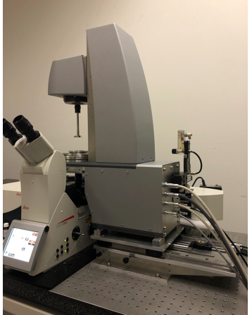
In collaboration with the groups of Prof Egelhaaf (Heinrich Heine University, Dusseldorf, Germany) and Prof Vermant (ETH, Zürich, Switzerland) we have constructed a novel high-resolution instrument, the confocal–rheometer, the first and only one in Australia
Confocal Laser Scanning Microscopy (CLSM) is a well-established tool for studying nanoscopic features and processes in cells and materials. However, this technique is often focused on fixed cell samples, and in cases of live cell imaging is limited to a static environment without the ability to dynamically change the matrix properties surrounding the cells.
The confocal–rheometer’s fine measurement capabilities enable applications in mechano-optical studies of synthetic ECM materials, where the influence of mechanical force applied on the material by the rheometer component can be concurrently visualized by the confocal microscope.
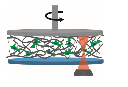
In the experiment shown in a schematic picture below, a soft polymer containing cells is visualized by fluorescence microscope while simultaneously applying strain to the sample by the rheometer plate on top.
We are also implementing this instrument in the study of various mechanosensitive processes in biological systems, such as focal adhesion formation kinetics as a function of externally applied force in 3D. We believe that this setup will provide us with new means of studying natural and synthetic ECM materials, understanding the processes associated with mechanotransduction in biological model systems and as an end goal to understand in more detail the interface of ECM and cells.